
Anesthetic considerations for robotic bronchoscopy
If you have not yet heard of robotic bronchoscopy, you are not alone


Figure 1. A Monarch robot bronchoscopy unit
I am joined in this post by Dr. Farhood Farjah, Professor of Surgery and Endowed Chair in Lung Cancer Research at the University of Washington.
I’m always fascinated by startling new things that I’ve never heard of. Recently a friend of mine announced that she was going to undergo robotic bronchoscopy for biopsy of a lung nodule. I had never heard of robotic bronchoscopy, and since I am a cardiothoracic anesthesiologist, I thought I had better look into what is going on. It turns out that this is a very interesting new technology and it has some specific considerations for anesthesia care.
Disclosure—I have no personal experience with robotic bronchoscopy, as our hospital does not currently have this equipment. Therefore my perspective reflects the available literature, which is fairly sparse, as this is new technology.
Bronchoscopic biopsy of lung nodules
Growing but small, part-solid or non-solid, or central lung nodules, discovered on CT scanning, require biopsy prior to resection or radiation. Non-surgical biopsy has taken two forms, transthoracic needle biopsy and bronchoscopic biopsy. The main drawback of CT-guided transthoracic needle biopsy is the high incidence of pneumothorax, around 30%. The main drawback of bronchoscopic biopsy is relatively low yield (false negative) results. Accuracy of bronchoscopic biopsy has been enhanced by navigational guidance such as endobronchial ultrasound or electromagnetic navigation bronchoscopy that uses a CT scan registered to the guidance system. In light of recent evidence of non-inferiority between transthoracic and navigational bronchoscopic biopsies in terms of accuracy, there remains an opportunity to improve the accuracy of non-surgical biopsies. This has led to the development of robotic bronchoscopy, the main benefit of which is more accurate navigation and biopsy tool delivery (Figure 1). Guidance is accomplished by a combination of registration to a previously obtained CT scan, fluoroscopy and other guidance technology, depending upon the particular robot utilized. There are currently 3 different robots that are commercially available. A very informative video describing robotic bronchoscopy can be found here.
Anesthetic considerations for robotic bronchoscopy
Robotic bronchoscopy for biopsies of small peripheral nodules typically requires general anesthesia. Moderate sedation may be possible and has been reported (see below) but is less likely to produce adequate conditions for these procedures which can be lengthy and require that the patients not move.
Atelectasis accompanying general anesthesia is an interesting issue to consider for these procedures. All navigational bronchoscopy requires pre-procedural CT scanning. The reference CT scans are taken at full inspiration. Differences between a virtual airway map generated from the CT scan and actual anatomy at the time of the procedure is known as CT-to-body divergence. Atelectasis and respiratory motion contribute to divergence.
Accordingly, there has been interest in using ventilatory parameters that might minimize atelectasis. Some recommended maneuvers include—
1. Avoid preoxygenation with 100% oxygen to avoid absorption atelectasis
2. Use the lowest possible FIO2 throughout the procedure to avoid absorption atelectasis
3. Tidal volumes of 10-12 ml/kg ideal body weight and PEEP 10-15 cmH20 (lesion in upper or middle lobes) or 15-20 cmH20 (lesion in lower lobes)—recommendation from Bhadra et al (retrospective comparison to standard care). R1 and R2 represent 2 different readers of CT scanning--“Atelectasis was more prevalent in the conventional ventilation group, both for dependent atelectasis (R1: 64% and R2: 68% vs. R1: 36% and R2: 16%, P= 0.00014) and sublobar/lobar atelectasis (R1: 48% and R2: 56% vs. R1: 20% and R2: 32%, P= 0.01). Similarly, the target lesion was obscured due to atelectasis more often in the conventional ventilation group (R1: 36% and R2: 36% vs. R1: 4% and R2: 8%, P= 0.01). Diagnostic yield was 70% for conventional ventilation and 92% for LNVP [lung navigation ventilation protocol] (P= 0.08)”.
4. As an alternative to the recommendation from Bhadra et al, the VESPA trial (randomized prospective trial) utilized tidal volume of 6-8 ml/kg of ideal body weight and PEEP 8-10 cmH20. “Patients in the VESPA group showed a lower incidence of intraprocedural atelectasis (any atelectasis, 28.9% vs 84.2% [P < .0001]” (atelectasis measured by CT scan).
How do these recommendations for ventilatory parameters during robotic bronchoscopy differ from “standard practice”? And what exactly is standard practice? “Lung protective ventilation” of patients in the ICU with ARDS with relatively small tidal volumes and PEEP to improve outcomes has generally been extrapolated to routine ventilation of patients with normal lungs in the operating room. A consensus paper recommended tidal volume of 6-8 ml/kg ideal body weight and 5 cmH20 PEEP for routine operating room ventilation. This is the same tidal volume as recommended in the VESPA trial for robotic bronchoscopy, but lower than the Bhadra et al recommendation of 10-12 ml/kg. The level of PEEP is lower than either VESPA or Bhadra et al recommendations. However, the evidence for the standard practice of 6-8 ml/kg and 5 cmH20 PEEP is not particularly strong. A systematic review and meta-analysis of intraoperative ventilation strategies by Bolther et al concluded--
“While our findings do suggest that some form of lung-protective ventilation (low tidal volumes with PEEP) decrease the risk of pulmonary complications, such a strategy did not translate into shorter hospital length of stay or lower mortality… Overall, there is limited evidence from randomized clinical trials to guide ventilation strategies during general anesthesia for adults undergoing noncardiac surgery.”
Bolther et al did not find differences in respiratory complications when comparing tidal volume of 6-8 ml/kg to 10-12 ml/kg. Likewise there was no difference in respiratory complications or atelectasis when PEEP>8 cmH20 was used in comparison to <5 cmH20. PEEP of at least 5 cmH20 did produce superior results to zero PEEP. Thus it appears that the VESPA trial recommendation of tidal volume of 6-8 ml/kg and PEEP 8-10 cmH20 for robotic bronchoscopy is reasonable and not greatly different from the recommendation for standard practice.
Moderate sedation for navigational bronchoscopy
Several studies have found comparable results for general anesthesia or sedation for ultrasound guided endobronchial biopsy. Casal et al performed a prospective randomized trial of sedation versus general anesthesia with a laryngeal mask airway for ultrasound guided bronchoscopic needle aspiration biopsy and found no difference in procedural success. However, it appears that robotic bronchoscopy is most likely to be performed under general endotracheal anesthesia. Despite the apparent preference for general endotracheal anesthesia there is a case report of successful use of the Ion robotic system under moderate sedation. They say a picture is worth 1000 words, see Figure 2.
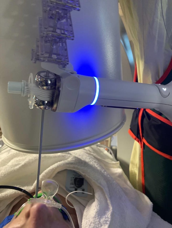
Figure 2. Patient undergoing Ion robotic bronchoscopy under moderate sedation. Robot arm above patient’s head. Photo from Donna et al.
If you have experience with anesthesia for robotic bronchoscopy I would like to hear from you. Please make comments and share your experiences with our readers.
 | A guest post by
|